Wednesday, June 29, 2011

Anatomy and Physiology Series: Laryngeal Physiology (Part 1, Say Yes to subglottal pressure--No to Bernoulli)

I'm going to split up laryngeal physiology into two posts because we've got two major things to discuss.  The first one, and the topic of this post, is the interaction of air pressure laws on vocal fold vibration.  The other, is the ideal interaction of the musculature to encourage efficiency.  I'm starting with air pressure laws because these are vital to understanding why the musculature must maintain such a delicate balance.  So here we go!


There are two main laws of air pressure we're going to talk about, the Bernoulli effect (which many folks speak of) and subglottal air pressure. 

We're going to go through the Bernoulli effect, or Bernoulli's principle, first, since it historically preceeded sub-glottal air pressure as an explanation for vocal fold movement.

Update (08/23/2015)
SCIENCE CORRECTION:  The Bernoulli Principle actually has nothing to do with vocal fold motion.  I know this is going against pretty much everything any of you have ever heard in your vocal pedagogy coursework, but the truth is that you only need the air pressure difference below the vocal folds (subglottal pressure) and above the vocal folds (supraglottal pressure) and the shape of the vocal folds themselves (the mucous lining in particular) to sustain vibration.  The issue with the idea of the Bernoulli Principle is that if that law really applied to vocal fold motion, the vocal folds would have no way to open back up again.  For all practical purposes for a singer, this doesn't really change much, but it is very important for those ENT's and researchers out there looking for ways to repair vocal fold scar tissue surgically and all that stuff.  The other nice thing about this as a singer (for me at least) is this: For all practical technical purposes, it's all about the subglottic pressure, which is great for us since subglottic pressure can be maintained through a lot of difference respiratory coordinations and configurations.  There might be an ideal out there for singers, but this, for me, explains why breathing is so important and such a variable, and sticky, pedagogical area for so many singers.

Subglottal air pressure is the measured pressure below the vocal folds.  This pressure and our intraoral (or inside the oral cavity) air pressure are pretty much equal to the pressure inside the lungs.  If we close the passage with our vocal folds, however, we instantly create an increase in the pressure below the vocal folds.  This is caused by the lungs continuing to exhale air which then "pushes" against that blockage (the vocal folds.)  While that is happening, the closed vocal folds also cause our intraoral pressure to drop to atmospheric pressure.  This increases the pressure difference between subglottal and intraoral pressure.  Now remember, air particles will always move from areas of high pressure to areas of low pressure, so subglottal pressure being significantly higher than intraoral pressure is a critical component to vocal function.  When this increased pressure exceeds a certain amount (3-5 cm H2O), the folds are blown open from the bottom, and the vibration known as phonation begins.* 

Here's how vocal fold vibration really works:  
The vocal folds are set into vibration through two mechanisms:  1. The glottal geometry (or shape of the glottis--the space between the vocal folds) and 2. The inertia of the vocal tract above the vocal folds.  As the subglottal pressure increases below the vocal folds, the vocal folds begin to separate at the bottom.  This is when the glottal geometry is convergent--when the top of the vocal folds are closer to each other than the bottom of the vocal folds.  In this configuration, the intraglottal pressure is high (that's the air pressure between the vocal folds themselves). This causes the vocal fold tissue to move away from the middle of the glottis.  But, vocal folds have elasticity thanks to that mucosa over the muscle.  This elasticity will generate a force that wants to restore the vocal folds to their original configuration.  This force will eventually overcome the force from the increased intraglottal pressure--this step happens from the bottom (inferior) vocal fold edges to the top (superior) edges and results in a rotational motion of the medial surface (middle of the vocal folds).  The vocal folds then take on a divergent configuration--the bottom of the folds are closer together than the top.  At the same time this rotational motion is happening, the intraglottal pressure decreases rapidly.  This causes the vocal fold tissue to change direction and move toward the middle of the glottis--this also happens from the bottom to the top.  The vocal folds either come together or get very close to one another (they don't actually have to make contact to create a sound wave--that's how you make breathy or lightly-voiced sounds) and then the glottis goes back to the convergent configuration and the cycle starts over again.  

From:  Titze, I. (1988).  The physics of small-amplitude oscillation of the vocal folds.  The Journal of the Acoustical Society of America, 83(4), 1536-1552.
The inertance of the vocal tract (#2 listed above) plays a part in this cycle in that as the vocal folds move outward from the middle of the glottis, the airflow through the glottis increases.  This makes sense, cause there's a larger glottis being created so more air particles can rush through.  This rush of air particles accelerates the air above the glottis that was previously relatively still. This increases the air pressure above the glottis (supraglottal air pressure) cause there's a higher number of air particles in that supraglottal space than there was before.  This increase of air pressure creates a force that moves the vocal folds outward.  This is where the elasticity of the vocal folds from above comes back into play, the vocal folds move back toward midline and the glottal flow decreases.  But, all those air particles that rushed through previously continue to move up the vocal tract because the inertia set those particles into motion and they want to stay that way until acted upon by another force.  This causes the pressure both above and within the glottis to decrease, allowing the vocal folds to come completely (or almost completely) together at midline and allowing the air pressure to build up below the vocal folds again.  Thus, the cycle continues as long as there's enough subglottal air pressure and continued air flow through the glottis at each cycle. *

So let's model this with a group of air molecules called Larry.  Larry gets sucked into the lungs during inhalation, he hangs out there a little bit, exchanges his oxygen for some CO2 and the forces of exhalation send him out.  But before he gets to go far, he encounters a blockage through his path.  This causes Larry to get smushed together with some of his other friends right under the vocal folds (like a little traffic jam).  Meanwhile, the air above the folds is decreasing in pressure, making it look like a really nice place to be.  The pressure Larry is under eventually overtakes the medial compression of the vocal folds, and the bottom of the folds begin to open.  Larry rushes through the vocal fold opening (unaware of the elasticity of the vocal folds cause, hey, he's just an air particle!) and then Larry runs into the air particles above the glottis and gives them a "push" to get them moving too.  As Larry and his air particle friends above the vocal folds travel up the vocal tract, the air pressure above and between the vocal folds decreases, the vocal folds come back together, another traffic jam starts up below them and the cycle repeats.  But Larry doesn't particularly care, cause he's already through the traffic stop.  A great visual model of this can be found at The National Center for Voice and Speech's website.  Scroll down for a more scientific explanation (certainly better than my "Larry" one) and some visuals that show this process.

The other nice aspect of taking out the Bernoulli concept is that it makes the concept of different vocal fold configurations for different voicing relatively simple to understand.  The vocal folds do not have to completely contact each other to create a sound pressure wave.  This is seen in regular voice users with videostroboscopy where you can see the vocal folds approximating during breathy or soft voicing and having a lot of contact during loud voicing.  It also explains something about the respiratory systems coordination for classical singing.  At high respiratory pressures, like when you've taken a really big breath, you are usually using high subglottic pressures to voice.  This increases the vocal fold contact time, resulting in greater swings of air pressures and producing louder (higher amplitude) sound waves.  Thus, vocal folds come together and stay together longer when voicing loudly. *

Another interesting aspect of this vocal fold vibration/air pressure relationship is that what is going on above the level of the vocal folds is just as important as what's going on below.  Therefore, if you have excess constriction above the vocal folds, you've changed these air pressure/air flow relationships that alter vibration and vocal fold contact times during vibration.  So, if you've ever had that "squeezing" feeling when singing, you likely had excess constriction above the vocal folds.  How to get that excess tension out is a whole different topic pedagogically, but I think it's enough to say that it requires a "resetting" of the air pressure/air flow relationships during that aria or difficult phrase, or maybe anytime you sing.  This will likely require some amount of "vocal play" between a new respiratory coordination (likely a bigger, lower breath), some amount of inspiratory checking (at least at the beginning of the phrase), and some concept that allows the supralaryngeal space (area above the vocal folds) to release the excess tension (like maybe the concept of an "open throat" or "singing on a yawn" or "inhaling a rose and singing with that space" or some variant of this).  

*Referrences:  

Hixon, T. J., Weismer, G., and Hoit, J. (2013).  Preclinical speech science: Anatomy, physiology, acoustics, and perception (2nd ed.).  San Diego, CA: Plural Publishing, Inc.

Raphael, L. J., Borden, G. J., & Harris, K. S. (2007).  Speech Science Primer:  Physiology, Acoustics, and Perception of Speech.  Lippincott Williams & Williams:  Philadelphia.

Titze, I. (1988).  The physics of small-amplitude oscillation of the vocal folds.  The Journal of the Acoustical Society of America, 83(4), 1536-1552.

Tuesday, June 28, 2011

Am I ever going to get back to telling my journey?

I had someone I know in my personal life ask me that the other day.  The reason I'm sharing this anatomy/physiology series with everyone is that it is a vital part of my journey thus far.  I recently had the pleasure to sing for a dear friend of mine who has heard my voice pre-therapy and in all stages of technical development post-stage, and who is a very talented, professional singer himself.  He agreed my voice has a much richer, fuller sound from top to bottom and that I have finally achieved a good "ringing" resonance in my middle voice, which is no small feat for a coloratura soprano.  


Now, I made a lot of gains in my singing before going through a year of undergraduate courses for SLP, but I must say, my largest gains have come from this year.  I haven't had many voice lessons at all this year due to time (note to self:  Do not plan a wedding while going to school full time while maintaining a full voice studio ever again!), and I certainly haven't been practicing as much as I'd like, so this breakthrough still isn't as consistent as it could be, but I have made huge gains in my singing.  How?  Learning in more intricate detail how the respiratory, laryngeal, pharyngeal, and articulatory systems work, in addition to some other things about how the brain works in terms of motor-development, etc., has really allowed me to take the concepts from my lessons that I never understood and play with those concepts during practice sessions, combining them with a great understanding of how it was all supposed to be working.  Not only that, but I can take a clear understanding of some biological functions and recognize those functions by a different name in pedagogical texts when I need another resource to turn to.  I have also become a much more effective teacher armed with this knowledge as well. 


In the current operatic world, young, professional singers are expected to have a solid enough grounding in technique to launch into a professional career at a young age.  At the same time, they are restricted to one-hour-a-week lessons either at the university, and sometimes, they get less time than outside of the university if their finances don't allow for once a week.  Yet, we expect them to not only arrive at the technical level of the singers of the past decades at a young age, but we expect them to maintain that level throughout their career with infrequent lessons scheduled between professional gigs.  Some singers do quite well with this model, and some are falling by the way-side.  Perhaps it's lack of talent, or perhaps some just don't get the "luck of the draw" in terms of getting in with a great teacher at a young age, but we can still do more to help all singers to succeed vocally with the current business model than we are doing (speaking of the operatic world in general terms here...there are pockets of great, intense training, but they aren't the norm).  


In the past, singers trained very differently.  They had a lesson nearly every day, rather than once a week.  Their training more closely resembled that of Olympic athletes in terms of personal attention, time, and training with the best coaches and teachers.  So they were able to make great debuts at young ages on the major opera stages of the world.  While the current financial situation many schools find themselves in don't allow for a return of that model, we do have more knowledge to offer our students.  We know so much more about the science of how the body and voice work, and what can go wrong.  Offering this knowledge to the students and singers of today shouldn't be seen as unnecessary or tedious, rather it should be seen as offering our students a safety-net with which they can save their own voice from harm while out on the professional circuit.  


Certainly, there are other things that lead to vocal demise, poor role planning, etc., but the signs of demise are so often overlooked by the singer, or are noticed, but the singer has no way of fixing the issues during the run of their show.  I personally believe that the most determined singers out there have the mental aptitude to learn about their instrument in more detail and to apply that knowledge to their own singing.  We expect singers to be so intelligent about so many things, languages, musicality, individual expression, yet we give them a pass when it comes to the detailed science of how the voice works.  Will this knowledge produce better singers?  Who knows.  But I bet it would to serve to protect some of the great unknowns out there, help them to separate the good pedagogy from the bad, and perhaps even expedite their vocal training.  


So that's why I'm going through this series.  I want everyone to have a resource through which they can understand how I went through my process and what helped me, including what I believe could help them as well.

Anatomy and Physiology Series: Anatomy of Extrinsic Laryngeal Musculature

So, we went through the intrinsic musculature on the last post, which are involved with the fine motor movements of phonation, including volume and pitch change.  Now, we're going to go over the anatomy of the extrinsic laryngeal musculature.  These muscles are divided into two groups:  Laryngeal elevators, and laryngeal depressors.  And that's pretty much what they do, raise and lower the larynx.  Hence, they're involved with the gross motor movement of the larynx.


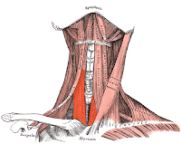
The main laryngeal elevators are:  Digastric anterior and posterior, the stylohyoid, mylohyoid, geniohyoid, genioglossus, hyoglossus, and thyropharyngeus muscles.  (Those last three muscles will be discussed in greater detail with articulatory and pharyngeal muscles.)  The Digastric muscle has two separate "bellies," or parts, the anterior and posterior.  (In red below:)


The anterior belly originates from the inner surface of the mandible (or lower jawbone) and the posterior belly originates from the mastoid process.  Both bellies insert into the hyoid bone at the top of the larynx.  The anterior belly pulls the hyoid up and forward and the posterior belly pulls the hyoid up and back.


The stylohoid muscle originates from the styloid process and inserts into the hyoid.  (Other than the diagastric, the other elevators have nifty names that help to remember origin and insertion.)  This muscle elevates and retracts the hyoid when contracted.
The mylohyoid muscle quickly became my favorite muscle during (timed) lab quizzes on laryngeal elevators because it's so easy to see!  This muscle originates from the side of the inner (or inside) of the mandible and fans inward to the hyoid bone, it's insertion point.  (This is really the primary muscle you're massaging when you stick your thumb under your chin...but if you really dig in, you could get at some of the lower tongue muscles as well.)  This forms the bottom of your oral cavity.  Contraction of this muscle moves the hyoid up and forward, and it also depresses (or lowers) the mandible.  It also raises the floor of your oral cavity when you're starting to swallow.
So the last elevator I'm really going to talk about in detail here is the geniohyoid.  The geniohyoid muscle is located directly above the mylohyoid, so when you look at the picture below, realize that the mylohyoid has been cut away so you can see it.  This muscle originates from the mental symphysis (which is the back of your chin) and inserts into, you guessed it, the hyoid bone.  (I told you that hyoid bone gets a lot of play.)  This muscle raises the hyoid up and moves it forward.
The other three, the hyoglossus, genioglossus, and thyropharngeus will be discussed in more detail when we get to the tongue and pharyngeal constrictors.  The first two listed, though, are primarily extrinsic muscles of the tongue, but they both elevate the hyoid as well.  The thyropharngeus is part of the inferior pharyngeal constrictor.  It has a bigger part to play in swallowing, but it does also elevate the larynx from it's insertion into the thyroid cartilage.  (Finally!  One that doesn't attach to the hyoid bone!)

On to the laryngeal depressors!  There are four of these bad boys we're going to talk about:  The sternohyoid, omohyoid, sternothyroid, and thyrohyoid.  I'll bet you $1 you know where their points of origin and insertion are right now.  (Seriously, though, never take a bet from a musician.  It's much more likely we actually don't have $1 to give you...)  Anyways, lets roll through this guys all lickity-split like, shall we?  (My apologies to my readers from outside of 1940's America.)

The sternohyoid originates at the sternum, more specifically the manubrium and the clavicle (now I'm just showing off) and inserts into the hyoid.  It lowers the hyoid upon contraction.  
The omohyoid (another one of my favorites during lab quizzes) has two bellies:  superior and inferior.  The two bellies are separated by the intermediate tendon, and as such, the superior belly originates from that tendon and inserts into the hyoid and the inferior belly originates from the scapula and inserts into the intermediate tendon.  Contraction of this muscle lowers the hyoid.
The sternothyroid originates at the sternum and inserts into the thyroid bone.  (Another one not hoping on that hyoid train.)  It lowers the thyroid cartilage.
And finally, the thyrohyoid, which originates from the thyroid cartilage and inserts into the hyoid.  (Yup, the hyoid is still a player.)  Contraction ends up lowering the hyoid bone, but it can also elevate the larynx if you talk to it really nicely.  (I'm getting a little punchy right now with all of this anatomy, if you can't tell.  It's just a little dry going through muscles that are named so clearly, but it does make life a lot easier on exams, let me tell you!)
There they all are!  The wonderful extrinsic laryngeal musculature.  This is the sling of muscles that suspend the larynx in the neck.  They have a whole lot to do with laryngeal posturing, or where the larynx is in the neck, and also laryngeal stability, which is a big deal for folks lacking muscle tone from a disease, disorder, or injury, but also useful for singers as well).  The delicate balance of coordination between the intrinsic laryngeal musculature and the extrinsic is a vital one to keep the articulatory system from trying to be the boss-man of the whole phonation-show.  

So what's up next?  The physiology of this whole system, including the vibration of the vocal folds (dominated by laws of air pressure) and laryngeal posturing during singing.  I might split it up into a couple of posts, so check back if I didn't cover everything in the next post.  

*Resource:  Seikel, J. A., King, D. W., & Drumright, D. G. (2010). Anatomy and physiology for speech, language, and hearing. Clifton Park, NY: Delmar.

Friday, June 24, 2011

Anatomy and Physiology Series: Intrinstic Laryngeal Musculature

Alrighty, now that we have a base for the underlying structure of the larynx, we can move on to the muscles that are part of making the whole thing work.  I'm going to talk about intrinsic muscles here, and I'll talk about the extrinsic in the next post.


So...intrinsic vs. extrinsic, what's the deal?  Back in the day when I was in vocal ped., we were taught that intrinsic muscles are all contained within the larynx itself.  That is, these muscles all have their origin and insertion points within the laryngeal structure (so somewhere on those cartilages we just went through.)  The extrinsic muscles have either their origin or insertion point in the larynx, but the other point is outside of it.
That is all absolutely true.  However, another important component of the difference between intrinsic and extrinsic is in their function.  Intrinsic muscles are concerned with fine motor movements and extrinsic are concerned with gross motor movements.  (This is good to remember, because the tongue also has intrinsic and extrinsic muscles with the same distinction.)   So for our purposes, intrinsic muscles will be involved in the minute vocal adjustments required for communication (pitch, volume, etc.), and the extrinsic muscles have more to do with where the larynx is located in the throat, i.e. elevated for swallowing, neutral for speech (ideally), lower for operatic singing (ideally) etc.  Also important for our purposes:  The intrinsic muscles are divided into adductors (or muscles that close the vocal folds) and abductors (muscles that open the vocal folds...there's only really one...I always thought it's sort of weird to have a whole category for one muscle), glottal tensors, and glottal relaxers.*


The first adductor is the lateral cricoarytenoid.  It originates from the superior-lateral surface of the cricoid cartilage, which is the top-front/side of the cricoid, and inserts into the muscular process of the arytenoid.  This muscle is a little hard to see in a model, because it is usually blocked from view by the thyroid.  (You can see it on the picture below because this view is from the side, as if the side of the thyroid had been cut away.)
When the lateral cricoarytenoid contracts, it pulls the muscular process of the arytenoid forward, rocking the arytenoid itself down and in.  This results in adduction of the vocal folds, and it may lengthen the vocal folds (but we're still not 100% sure about that one).


The transverse arytenoid muscle originates from the outer side of the arytenoid and inserts into the outer side of the other arytenoid.  Contraction results in pulling the two arytenoids closer together than just the lateral cricoarytenoid by itself.  This muscles as a lot to do with medial compression, which is the degree of force that brings the vocal folds together at the midline.  It's the muscle underneath the criss-cross muscles pictured below.


Those criss-cross mucles above are the oblique arytenoids.  They originate from the posterior base of the muscular process of the arytenoids (so...the back-bottom of the arytenoids) and insert into the top of the opposite arytenoid.  Contraction results in pulling the top of the artyenoids towards the midline, which grants stronger adduction through medial compression, and rocks the arytenoids down and in (so it supports and adds to the motion of the lateral cricoarytenoid).


The abductor of the vocal folds is the posterior cricoarytenoid muscle, and you can see it in the above picture as the lowest muscle pictured.  This guy originates from bottom-back of the cricoid and inserts into the bottom-back of the arytenoid (so it courses upwards).  Contraction results in pulling the muscular process of the arytenoids back, which rocks the arytenoid cartilage outwards and abducts the vocal folds.  I believe scientists used to think this muscle didn't have a whole lot to do...which makes sense given muscular elasticity would naturally abduct the folds upon the end of phonation, but we now know that this guy has a big part to play during speech and inhalation.  This is what allows for more opening of the glottis when we're breathing, either during exercise or at rest, and it also opens the glottis when we make plosive consonants that require very fast, short bursts of air through the larynx.  So it's actually a pretty active little muscle...and loss of function in this guy causes a lot of issues for folks being able to breath easily.


The two glottal tensors are the cricothyroid muscle and the thyrovocalis muscle (which is a part of the thyroarytenoid muscle).  The cricothyroid actually has two parts to it:  the pars recta ("straight part") and the pars oblique ("angled part").  The pars recta originates from the front surface of the cricoid and inserts into the lower part of the back-front of the thyroid.  The pars oblique originates just to the side of the pars recta origination, and inserts higher up on the thyroid cartilage than the pars recta.  The pars recta portion is what rocks the thyroid cartilage downward.  (This movement does result in the cricoid rising, but that is due to the flexibility of the tracheal cartilage rather than direct muscular movement.)  This movement stretches the vocal folds, changing the pitch of the voice.  The pars oblique portion slides the thyroid slightly forward (via the thyrocricoid joint), which also tenses the vocal folds, elevating the vocal pitch.  (Unfortunately, I couldn't find a good, public-domain image of this muscle for ya.)


The thyrovocalis is the middle muscle of the vocal folds.  In fact, this is the muscle that makes up the inner layer of the vocal folds.  It originates from the inner surface of the thyroid cartilage and inserts into the vocal process of the arytenoids.  Contraction of this muscle tenses the vocal folds by drawing the thyroid and cricoid cartilages farther apart.  Notice that that motion is the opposite of the cricothyroid muscle, which pulls the thyroid and cricoid together by rocking and sliding.  Although it moves the thyroid and cricoid in the opposite way of the cricothyroid muscle, it works in conjunction with it to tense the vocal folds even further through the antagonistic movement.  (For yogis out there:  This is a bit like how drawing the arms up into the shoulder blades while simultaneously lowering into "push up" position results in more muscle tone to support your body weight...sort of.  Can't think of a better example right now...)


The glottal relaxer, the thyromuscularis, is paired with the thyrovocalis in a special way I'll talk about below.


This muscle originates from the same place as the thyrovocalis and inserts into the muscular process of the arytenoids.  Contraction of the more-medial (or middle) portion of this muscle also has the same results as the thyrovocalis.  However, contraction of the the same section might also relax the vocal folds by pulling the arytenoids toward the thyroid cartilage without changing the rocking motion of the thyroid.  So, anatomically, there is not really a difference between the thyrovocalis and the thyromuscularis, but functionally, they are distinctly different.  Hence, some sources lump them together into the thyroarytenoid muscle rather than maintaining a distinction in function by separating them.  However, since operatic singing requires more finely-tuned laryngeal function than daily voice use, I feel this distinction is an important one to highlight.


And there you have it.  Your intrinstic laryngeal muscles (or the main ones we'll talk about anyway.  There are some supporting muscles to these, but their impact on vocal function is questionable, while their necessity in the swallowing process is well known.  I'm just not going to get into swallowing here.)


*Citation:  Seikel, J. A., King, D. W., & Drumright, D. G. (2010). Anatomy and physiology for speech, language, and hearing. Clifton Park, NY: Delmar.

Anatomy and Physiology Series: Laryngeal cartilages (and hyoid bone too)

Now we get to the fun stuff...okay maybe not quite yet.  This is more like the dry anatomy build-up to the fun stuff (laryngeal physiology).  Much of what I say here can be found in pretty much any vocal pedagogy book out there, but I think it's still important to go over.  Even if you feel you know this from a pedagogy course you took in school, the information is worth reviewing, and who knows...you might find there's something new in here as well.

Let's start with the 5 main cartilages (and hyoid bone) of the larynx.  These cartilages and one bone make up the internal structure of the larynx and serve as attachment points to the intrinsic laryngeal musculature.  The 5 main cartilages are:  The thyroid cartilage (which the thyroid gland sits on the bottom of), the cricoid cartilage, the two paired arytenoid cartilages, and the epiglottis.  (There are other cartilages, the corniculate and cuneiform cartilages, but they don't have quite as much to do with voice production.)  I'm including two pictures below of these structures.  In one, they're all separated, and in the other, they're all together as they are in the body, as viewed from behind.


The thyroid and cricoid cartilages are attached by the cricothyroid joint (or articulation as in the above picture), which allows for the "rocking" motion of the thyroid during phonation.  The epiglottis is considered the entrance to the larynx.  It mainly serves to close off the glottis when swallowing (by folding down when the larynx elevates), but it also forms part of the pharyngeal wall during phonation.


The hyoid bone does not directly articulate to any other bone in the body.  However, it is attached to the thyroid via the hyothyoid membrane.  Why is it a bone and not cartilage like everything these?  I'm not sure, but I assume by being a more sturdy material, bone, it provides stronger support for the laryngeal cartilages.  The hyoid bone is a major point of attachment for many muscles above the larynx.  I believe it has something like 30 or 40 muscles that attach to it (the actual number escapes me at the moment).  We're only going to talk about a few, cause we're much more interested in the laryngeal elevators and articulatory muscles.


I do want to discuss the arytenoids in some detail here before getting into laryngeal musculature.  The arytenoids are pyramidal-shaped cartilaginous structures (and from a certain angle, they sort of look like a shark's tooth.)  The joint that attaches them to the cricoid allows for rotation of the arytenoids, which allows the vocal folds to close.  There are two main processes (or protrusions) that should be discusses here.  The first one, the vocal process, cannot be viewed from above (unfortunately), but it is the point near the base of the arytenoid on the opposite side from the view you currently see up there.  The other, the muscular process, can be seen.  It's the point at the base on the outside of each arytenoid in the above view.  These two points serve as important attachment points for intrinsic laryngeal musculature, so it's good to remember them.


Okay, so I was going to launch right into intrinsic laryngeal musculature, but I think if I did that, this would turn in to some monster-post.  So I'll just finish this one here and start up with intrinsic musculature next.

Thursday, June 16, 2011

Anatomy and Physiology Series: Physiology of Exhalation (and breath support)

You may have heard that we don't really use all the air our lungs take in. This is very true.  In fact, pulmonologists and other folks concerned with the health of the respiratory system have come up with several volumes to determine healthy lung function.  


Let's go over some of these volumes so you'll have some idea of what I'm talking about when I refer to them:  Tidal volume (TV) is the volume inhaled and exhaled during one cycle of respiration.  It varies from resting to exercise and age of person, etc.  Inspiratory reserve (IRV) is the air inhaled beyond the tidal inspiration.  Expiratory reserve (ERV) is the air expired beyond tidal expiration.   Vital capacity (VC) is the one most often referred to in voice research.  VC is the volume available for speech.  It is a combination of IRV, ERV, and TV.  (Others we won't talk about are:  Residual volume (RV) is the air that remains in the lungs after maximum expiration...it's always in there.  Functional residual capacity (FRC) is the air that remains in the body after passive exhalation, inspiratory capacity (IC) is the volume that can be inhaled from resting lung volume, and total lung capacity (TLC)  which is the sum of all lung volumes.)*


During respiration, the forces of exhalation are passive if we stay above 38% of our VC. However, when we speak professionally and sing, we have to maintain fairly consistent subglottal pressure (which we'll get to in detail later) while going beyond that 38%.  Not only that, but we have to slow our exhalation prior to getting beyond that 38%.*  So how do we do all of that?  By using a combination of our checking action combined with our musculature of forced exhalation.


Checking action* is the process by which you check (or hold back) the flow of air out of your inflated lungs by means of the muscles of inhalation.  When singers heighten this and expand it to singing, they call this the appoggio technique.  It is a natural occurrence during speech, and is especially noticeable during public speaking.  (It is no wonder those pedagogs of the "golden age of singing" came up with this as a key to proper singing.  It's a coordination that is already there!)  It's built into the healthy-functioning adult body by the biological respiratory system responding to the communication demand the brain is putting on it.  Singers need to improve this coordination, making it the most effective possible and using even more muscular energy to sustain even longer phrases of communication (i.e. music.)  I have found that training this natural coordination will inherently involve a student going a little too far with the coordination, either resulting in too little air flow into the throat and/or too much air flow, but I think that is a natural process of heightening this coordination to meet the demands of singing.  Once a singer acquires this more precise coordination, it really just happens "in the background" of his/her brain as long as they actively maintain the muscular strength through constant usage (ala the professional singer with consistent practice habits.)  One of the abilities of a great teacher is to know when a student needs more strength/coordination training and when that student needs to back off of conscious control of the coordination to allow it to become "second nature."  (If you don't ever "back off," I believe you are in danger of eliciting a different coordination discussed below.) 


Exhalation musculature is actually only used when expiratory reserve needs to be tapped into.  When you get below the 38% point of VC during speech/singing, your body will call on its muscles of forced exhalation to push beyond that point so that we can keep right on going.  This action occurs sequentially along with the checking action to utilize all of your expiratory reserve before your next breath, if necessary.  


This coordination of checking action, with forced exhalation taking over at the appropriate point, takes a lot of muscular effort when sustained for a long period of time...as when the teacher lectures for an hour, the actor performs a two-hour play, or the singer sings for the two hour opera.  That is why we need to train in the strength and endurance for this coordination!


However, within our training there is a biological function that we need to be aware of so that we can counter it's effects.  This is called abdominal fixation* and it is the process of holding air in the thorax to stabilize the torso.  (This is what results in "grunting" when you're lifting that heavy couch while helping your friend move.)  It is a biological function that occurs when the body needs more rigid support and comes from the action of abdominal contraction along with full glottal closure to "hold" the air in the lungs.  It is the action that makes your chest and torso inflexible for more strength.  (This is also why your exercise instructor will remind you to "breath" during weight lifting and/or abdominal crunches and/or strenuous workouts.)  If you're brain tells your torso you need some major "support" for the task at hand, this is the coordination that your brain will naturally tell your body to do.  Obviously, closing off the laryngeal opening completely does not bode well for the singer, and it is exactly this motion that, I believe, lends itself to "pushing" the voice.  How do we counter it?  Well, I think a conscious realization that, since we're trying to maintain continuous breath flow, it must be a system that is capable of moving.  It must be able to make minute* adjustments to our air flow upon command, and therefore, it must retain a certain amount of flexibility.  It's not an abdominal "crunch," it's a belly-dancer's stomach "roll"...sorta-kinda.


So how do we train good breath support for singing?  I believe it must be based in the natural support mechanism we already use for sustained speech.  We must strengthen that mechanism to build our endurance and checking action while not encouraging the abdominal fixation of "rigid" support to creep in a close off our laryngeal system altogether.  So this is where we get the idea of strong, yet flexible support.  Some aspects will be rigid:  Such as our inhalation muscles of the upper back and our exhalation muscles of the lower back.  These guys will stabilize the torso while the other muscles of inhalation and exhalation do their work of checking and forcing exhalation at the appropriate moments.  It is a delicate balance, it is incredibly frustrating to figure out, but if you accept that it is not a completely foreign bodily function, then it might just work itself out faster than you think. 


*Just how minute the changes are that are required will be gone over during the laryngeal physiology.  So in the meantime:  Prepare to have your mind blown!  (Maybe...well...it blows my mind at least!)*


*Citation:  Seikel, J. A., King, D. W., & Drumright, D. G. (2010). Anatomy and physiology for speech, language, and hearing. Clifton Park, NY: Delmar. 

Anatomy and Physiology Series: Anatomy of Forced Exhalation

As discussed earlier, expiration is mainly the result of passive forces, but during speech (and singing), expiration requires more muscular effort.  This is referred to as "forced exhalation."  I'm going to talk a lot about the physiology in the next post, but first, let's get ourselves grounded in what muscles are involved.


The first group I'll introduce are...*drum roll*...the abdominal muscles!  This should come as no surprise to the experienced singer, but let's still go over where they are and what they do.  The abdominal muscles as a group are involved with changing the vertical dimension of the thorax through affecting the movement of the diaphragm.  (It'll go lower with relaxed abdominal muscles, and higher, faster with contracted abdominal muscles on the exhale.)  If you've ever wondered what the abdominals attach to, I would recommend reading about the abdominal aponeurosis.  (I'm not going to cover that here in the interest of space.)  So here are the muscles we'll go over: The internal and external oblique abdominis, transverse abdominis, and the rectus abdominis on the anterior (front) part of the body.  The posterior (or back) ones include the quadratus lumborum iliacus and psoas major and minor.  (Some say the latissimus dorsi muscles are also involved, but there is currently some doubt to this claim, so I'm excluding it here.)  I'm going to cover these muscles in this order:  Anterior first, going from the deepest muscle (furthest in the body) to the most superficial; then, we will cover the posterior muscles.


The deepest and most powerful abdominal muscle is the transverse abdominis.
This muscle originates from the posterior abdominal wall at the vertebral column and inserts into the abdominal aponeurosis and the inner surface of ribs 6-12.  It is here, at ribs 6-12 that this muscle, here's a fancy term for ya, interdigitates (or interconnects) with the fibers of the diaphragm.  It's lowest attachment is the pubic bone.  Contraction of this muscle significantly reduces the volume of the abdomen.  (It's the "sucking in the tummy" muscle, and it's a pretty powerful one.  Not the origin of your desired "six pack," though, since it's too deep in the body to see outlined through the skin.)


Next, the internal oblique abdominis, and it's between the external oblique and the transverse abdominis.  
It's origin is from the iliac crest and it inserts into the cartilaginous portion of the lower ribs.  If you want to rotate your trunk, this muscle contracts on one side of the body, but if you contract both sides, it flexes the trunk ("curls" your back) and compresses the abdomen.  


The external obliques are the most superficial of the abdominal muscles, meaning they are the closest to your skin.  They are also the largest of the abdominal muscle group (but not the most powerful, that's still the transverse abdominis).  


Contraction of these guys can rotate the trunk as well and can compress the abdomen and vertebral column by contracting both sides.  


The rectus abdominis muscles are the "six pack abs" group.  (You can see them as the "rectangular" muscles in white in the picture above.)  Contraction results in flexing the vertebral column.


The first posterior abdominal to discuss is the quadraus lumborum pictured in red here:
These muscles originate from the iliac crest and insert into the lumbar vertebrae and rib 12.  It assists you if you move your trunk laterally (ala salsa dancing), and contraction on both sides fixes the abdominal wall in support of compression (ala my yoga instructor's directions for a good hand stand...still working on that one).  


Psoas major and minor are relatively smaller muscles hanging out around the pelvic area.  If they assist with exhalation, they are similar to the quadratus lumborum.


Other muscles of forced exhalation are mainly involved in decreasing the size of the rib cage.  These include the internal and innermost intercostals, transversus thoracis, subcostals, and serratus posterior inferior.  


The internal intercostals are deep to the external intercostals and their fibers run perpendicular to the external intercostals.  These guys depress ribs 1-11.  The innermost intercostals are actually a separate group.  There are the deepest of the intercostal muscles.  They run parallel to the internal intercostals and also depress ribs 1-11.  

The transverse thoracis originate at the sternum and insert into the inside of the rib cage.  This muscle group also depresses the rib cage...big surprise, huh?  Shown in red below (the sternum is seen on your right):

Posterior thoracic muscles include the subcostals.  These guys run similarly to the internal intercostals, but they can span more than one rib depending on the person.

The serratus posterior inferior originate from the spinous process of thoracic vertebrae #11, 12 and lumbar vertebrae 1-3.  They insert into the lower five ribs and also aid in pulling the rib cage down.
If you're still awake, congratulations!  You've now entered full-on voice-science geek status.  (just kidding).  But seriously...although I won't go so far into detail during the physiology discussion, I believe it is very important to not only understand how inhalation and exhalation work from a biological standpoint, but it is also important to know how complex the act really is.  It involves a very complex muscular coordination that, for the most part, all goes on in the "background" of a person's brain function.  It is because this process usually happens automatically for most people during speech that I believe the training of "breath support" can become an incredibly frustrating endeavor for the professional voice user, and it can be equally frustrating for a voice teacher to explain in a 60 minute lesson.  

The singing world is full of many "schools" of breath support and navigating those schools can be very frustrating for the student of pedagogy.  I believe having a more thorough knowledge of the known functionality and coordination of breathing for speech can be incredibly useful when navigating the more abstract ideas for breath support.  

So there's my soap-box for today!  Onward to physiology of exhalation!


*Re-citing:  Much of my material is coming from:  Seikel, J. A., King, D. W., & Drumright, D. G. (2010). Anatomy and physiology for speech, language, and hearing. Clifton Park, NY: Delmar. 

Wednesday, June 15, 2011

Anatomy and Physiology Series: Physiology of inhalation

Inhalation gets pretty largely ignored in some pedagogical circles...not all, but some. Singers tend to be all about the exhalation:  How to make it longer, how to make it easier, how to make it stronger, but they very rarely take a look at inhalation. At rest, inhalation is mainly done by the contraction of the diaphragm. When breathing at rest, inhalation is about 40% of the breathing cycle, and exhalation is about 60%. When speaking, inhalation becomes about 10% of the cycle and exhalation about 90%. And, although there is no solid data on this, I would suspect during operatic singing, that gap is even wider between inhalation and exhalation. So let's go over the two main ways a person can inhale for speech and singing and their biological function so we can get a better sense of how to make that 10% of our breathing cycle the most efficient for our use.

The first, ideal inhale is the one that yields the maximum expansion of the thorax. The most effective way to do this is to maximize the descent of the diaphragm and illicit the use of the primary rib cage elevators (more than just the intercostals) during inhalation. The first part is where relaxation of the abdominal muscles come into play. If the belly relaxes out, the internal viscera (otherwise known as your guts) move out with it, creating more space for the diaphragm to descend into. The second part makes use primarily of the external intercostal muscles, the interchondral portion of the internal intercostals, levator costarum, and serratus posterior superior. These guys all work together to obtain the maximum expansion of the thorax in the most efficient way for speech. (If you take part in "chest" breathing, you're mainly only using the chest expansion without the diaphragm descent, so it just isn't as effective as it could be.)  

The accessory muscles of the neck, mentioned in the second half of my last post, come into play either when you're really needed some extra oxygen, like on the last leg of that marathon you're training for, or during clavicular breathing.  This is associated with some pretty bad medical conditions, like COPD and neuromuscular disorders like ALS, but anyone whose had an asthma attack, anxiety attack, or ever had pneumonia have done a bit of clavicular breathing themselves. This gets the accessory muscles of the neck and back involved. In fact, in extreme cases of respiratory distress, patients tend to hold onto backs of chairs to try to get the pectoralis major to expand the rib cage. (Since that is not the primary function of that muscle, it's considered a desperate move for the body to gain a little extra oxygen.) This is considered the most inefficient way for the body to breath, so why does the body do this if it's so inefficient? Well, consider what happens to folks having an asthma attack where the airflow is obstructed. If the lungs cannot properly expand, then the muscles of inhalation won't really work either, thanks to the relationship between the muscular function and the difference in air pressure.

Now the thing is, it's not just in clavicular breathing that those accessory muscles of the neck can get involved, it is any time your body needs that extra oxygen elevating the rib cage affords.  Anyone who's done any intense exercise  can tell you that the body basically uses a combination of all three of types of inhalation to get every bit of air you need.  Ever worked out in the morning and gone into the practice room in the afternoon and noticed your neck muscles were tight?  It's very likely they were working during your work out too.  But, in singing inhalation, we don't want the neck muscles to be involved, so I feel it is always a good idea to massage out those muscles after a workout before you practice to make sure they're not getting in the picture.

High chest breathing comes into play biologically when we're in a heightened state as well, such as the fight or flight response.  This is another reason a low, relaxed inhale should really be well-trained into the singer's technique.  Those performance nerves can easily cause the body to tap into the wrong muscular balance.  I'll get into that a bit later down this series, mainly when I talk more about the nervous system physiology, but I feel it warrants being mentioned here that the body has this ingrained, biological responses to certain stimuli.  Nervousness and anxiety can trigger the body to get the higher, clavicular and/or chest type of breathing involved when we don't want it to be.  It doesn't mean you have bad technique inherently, but it does mean you need to work on what triggers that response and try to counter it either with more muscular training, i.e. muscle-memory, or with relaxation techniques.  Whichever works for you!

Another thing I will get into more after going through exhalation anatomy and physiology is the laryngeal system's role in the respiratory system.  But keep in mind until we get there:  The laryngeal system is a protective system biologically.  It is primarily a valve designed to keep food out of the lungs.  If you suffer from the "noisy inhale" issue some of us face at one point in our development, try slow, low, relaxed inhales through an open throat.  There should be absolutely no sound, even to you, as you breathe this way.  It is very possible that if you use too much of the accessory neck muscles during inhalation for singing, you are triggering the valve-effect of protection while you're gasping away for air.  (I'm not sure as to why, but I suspect it could have something to do with wanting to protect you from inhaling large objects, like flying insects, into your lungs when breathing rapidly through your mouth.)  But if you know that it's a biological function, it's easier to discover how you will insight relaxed breath for yourself through a little trial-and-error in the practice room.


*Citation: Seikel, J. A., King, D. W., & Drumright, D. G. (2010). Anatomy and physiology for speech, language, and hearing. Clifton Park, NY: Delmar. 

Tuesday, June 7, 2011

Anatomy and Physiology Series: Inhalation Anatomy


I've been reading up in my handy-dandy textbook from the previous post to try to figure out just what is the most important stuff to include for students and teachers of singing, as well as anyone making public speeches. However, I have encountered the issue of an abundance of riches: There's so much information that I think it'll be too hard to include it in just one post on inhalation and one post on exhalation as I planned. So, I'm going to split up some of these posts, where I see fit, and do inhalation anatomy as one post and some key inhalation physiology as another post before moving on to exhalation. I feel there is so much contradictory "theory" of breath support in the singing world out there that an effort to explain more scientific detail than what is usually gone over will be worth it...if I pull it off.


Inhalation can be an over-looked issue with some singers out there. I work a lot with inhalation when I get students who need the work. This is due to the ways in which a person can inhale, i.e. the many different coordinations of the musculature of the torso, thorax, and neck, and the biological function of those different coordinations.  We'll get into those functions in the next post, but for now, on to the inhalation musculature!

We have all heard, in one way or another, of the primary muscle of inhalation, the diaphragm.  This is the bowl-shaped muscle that rests under the lungs.  It flattens downward upon contraction (inhalation), and returns to it's resting bowl-shape on exhalation thanks to muscular elasticity.  It is innervated by the phrenic nerve, which originates in the cervical plexus.  It is bilaterally innervated, meaning there is nerve routes to the diaphragm on both sides of the body.  This is mainly a protective measure so that if there's damage on one side, the nerves on the other side will keep it working (a sign that it's a pretty important muscle for the body.)  It is attached to the lungs via pleural lining and the central tendon, which is the big white area on the picture below.  When you're at rest, this is the main muscle controlling inhalation.  (note, this is an inferior view, basically looking up through the abdomen.  The central tendon, however, is on the superior (top) of the diaphragm, so they have it here just to show it.  In fact, the heart is above the central tendon.)

These other muscles of inspiration are considered accessory muscles.  They are considered such because if you're knocked unconscious, this muscles aren't really used for inspiration at rest.  (Physics and elasticity from the last post does the work there.)  So these guys help mostly in what we call "forced inspiration," like what we do when we speak, sing, exercise, etc.  The external intercostals are included in this category (although they do play a role in resting inhalation as well, we could breathe at rest just fine without them...so they're considered accessory as well.)  These guys run between the ribs, are "external" to the internal intercostals (expiration muscles) and do not attach all the way around the ribs, stopping short of the sternum because attaching there would not do any good in elevating the ribcage, which is what they do.  You can see them labeled between the first and second rib show in this picture.  (That large, flat muscle shown here is one of the internal oblique abdominals...we'll get too on expiration.)


The other accessory muscles of inspiration, like the intercostals above, have to do with elevating and expanding the ribcage in some way.  These include the levator costarum, located at the back of the ribs near the spine.  There are 12 pairs of these muscles for each of the 12 ribs.  (They're the little red guys in the image below.)


The serratus posterior superior are paired muscles that originate from the spinous process (part of the vertebrae that sticks out) and inserts into the upper borders of the 2nd through the 5th ribs.  I'm having a heck of a time finding a good image that I trust won't carry some virus or something (been burned by that before!), so I'll just link to an image I like here:  They're the group labeled #1.  

There are other accessory muscles I have deemed less important for the singing/teacher/speaker to know, but I'll list them along with a link if you want to know more:  erector spinae, lateral bundle, and intermediate bundle.  These guys are more involved in spinal and rib cage stabilization than respiration directly.  (The last two in that list are on the first link.)

But, we're not done yet!  If you're not totally asleep by now, (and if you are, I don't blame you, anatomy without good physiology to back it up is pretty darn dry!), I'm gonna add in some of the accessory neck muscles to respiration.  These are primarily the sternocleitomastoid and the scalenes.  (If you've taken a pedagogy class before, you've probably heard of at least one of those, huh?)

The sternocleitomastoid is a strong muscle of the neck.  It originates from the mastoid process (the bump you feel right behind your ear) and has two points of insertion, the sternum and the clavicle (put all three together, and you've got it's name).  If just one side of this muscle is contracted, it results in rotating your neck towards the contracted side.  If both sides are contracted, it results in the sternum and clavicle (and by association the upper rib cage) being elevated.  But I like to think this muscle is the most happy when it's busy rotating your neck instead of aiding in respiration.


The scalenes (anterior, middle, and posterior) is a group of three muscles that, biologically speaking, help to stabilize the head and help out in neck rotation as well.  But, since they also insert into the 1st (for the anterior and middle scalenes) and 2nd (for the posterior) ribs, they can also elevate the upper rib cage.  (In the image below, you can see the anterior and middle scalene under the sternocleitomastoid.  The posterior one is being "hidden" by some muscles that go over it.)


Are we done yet?  (That's what I'm thinking right now, but no, I'm not done yet...I suppose you could be, though, since this is a blog entry after all.)  There are muscles of the arm and shoulder that aid in forced inhalation as well.  I think I'm just going to link to some of them, cause some are known pretty well in pop culture thanks to some hollywood hotties out there.  Pectoralis major and minor both elevate the sternum, which ends up increasing the rib cage in transverse (or side-ways) motion.  Serratus anterior help to elevate ribs 1-9.  Levator scapulae, which is actually what's hiding the posterior scalene in the image above, is a neck stabilizer, but it can elevate the rib cage as well.  

I'm going to include these next guys here because I feel they are key players in proper breathing posture for operatic singing (but, keeping this entry strictly dry, boring, and all about the anatomy).  Rhomboideus major and minor are often included in inspiration since their main function is to stabilize the shoulder girdle.  The trapezius is also often included.  This guy is a pretty big, strong muscle.  It supports arm movements, moves (and can stabilize) the scapulae (i.e. shoulder blade), elongates the neck, and helps to control head movement.  (You might have found yourself working this one out in the gym, along with pectoralis major.)  


So what do you have to look forward to with inhalation physiology?  The difference between diaphragmatic breathing and clavicular breathing, biological functions of both of these, and which of these muscles we just went over are our friends and which ones are our enemies when it comes to singing...duh, duh, duuuuuuuhhhh!!  (That's my poor attempt at suspenseful-sting music there...yeah, it's time to go.)


*Citation: Seikel, J. A., King, D. W., & Drumright, D. G. (2010). Anatomy and physiology for speech, language, and hearing. Clifton Park, NY: Delmar.